Knockout Cell Lines

Home > Gene Editing Services > Stable Cell Lines > Knockout Cell Lines

Knockout Cell Line

According to the characteristics of different cell lines, appropriate transfection methods (virus transduction, liposome transfection, and electroporation) will be selected to transfer gRNA sequences and Cas9 protein into cells, and antibiotic screening with different duration will be carried out according to different transfection methods used. After antibiotic screening, single-cell clones will be generated. Positive clones that are successfully knocked out will be validated by target site amplification and sequencing. Final deliverables will be the homozygous KO cell clones, related data and project reports.
KO cell bank
 3000 KO cell lines    Starting from $1780    Deliver in week!  

Ubigene's KO cell bank covers thousands of genes, including 8 signal pathways, 10+ drug development sectors, 100+ common diseases and popular research fields (such as m6A, ferroptosis and exosomes). Search your target gene below to find out if there is a KO cell line in stock.

Knockout Cell Service
Cell typeVarious types of cells including tumor cell lines, regular cell lines, IPS/ES cell lines
Service typeSingle / Multiple Genes Knockout
DeliverablesCell pool/Single-cell clone
Turnaround/PriceSpeedy turnaround as fast as 4 weeks!     Contact Us
Click to view articles citing Ubigene

200+Successful
Gene-editing Cell Line Types

Respiratory System
+
Chinese hamster lung cells(V79)Human hypopharyngeal carcinoma cell line(FaDu)Human Bronchial Epithelial Cell Line(16HBE)Human Bronchial Epithelial Cell Line(BEAS-2B)Human Non-small Cell Lung Carcinoma Cell Line(HCC827)Human Non-small Cell Lung Carcinoma Cell Line(NCI-H1299)Human Lung Squamous Cell Carcinoma Cell Line(NCI-H226)Human lung squamous cell carcinoma cell line(SK-MES-1)Human Lung Cancer Cell Line(NCI-H520)Human Lung Cancer Cell Line(Calu-1)Human Lung Cancer Cell Line(A549)
Circulatory System
+
Mouse Myoblast Cell Line(C2C12)Human Coronary Artery Endothelial Cell line(HCAEC)Rat Cardiac Myocytes(HL-1)Rat Cardiac Myocytes(H9C2)Human Umbilical Vein Endothelial Cell Line(HUVEC)
Endocrine System
+
Human Breast Cancer Cell Line(MCF7)Mouse insulinoma β cell line(NIT-1)Human Breast Cancer Cell Line(JIMT-1)Human breast cancer cell line(T-47D)Human pancreatic cancer cell line(BxPC-3)Mouse Acinar Pancreatic Cell Line(266-6)Human Prostate Cancer Cell Line(VCaP)Human Pancreatic Carcinoma Cell Line(MIA PaCa-2)Mouse medullary breast cancer cell line(E0771)Mouse pancreatic cancer cell line(Pan02)Human Metastatic Pancreatic Adenocarcinoma Cell Line(AsPC-1)Human Breast Adenocarcinoma Cell Line(SK-BR-3)Human Pancreatic Carcinoma Cell Line(PANC-1)Rat Breast Cancer Cell Line(4T1)Human Breast Cancer Cell Line(ZR-75-1)Human Breast Cancer Cell Line(MDA-MB-231)
Brain and Nervous System
+
Human glioma cell line(U251MG)Mouse microglia cell line(BV2)Immortalize Human Microvascular Endothelial Cell Line(hCMEC/D3)Mouse Anterior Parietal Bone Cell Line(MC3T3-E1 Subclone 14)Human glioblastoma cell line(U-87 MG)Rat Glioblastoma Cell Line(C6)Mouse neuroblastoma cell line(Neuro-2a)Human Neuroblastoma Cell Line(SK-N-SH)
Blood and lymphatic System
+
Human B lymphoma cell line(Su-DHL-4)Human B Cell Lymphoma Cancer Cell Line OCI-LY3(OCI-LY3)Human B Cell Lymphoma Cancer Cell Line(U2932)Human Acute Non-B Non-T Lymphocytic Leukemia Cell Line(Reh)Human myelogenous leukemia cell line(K-562)Human T lymphocyte cell line(Jurkat, Clone E6-1)Rat Basophil Leukemia Cell Line(RBL-2H3)Human Monocytic Cell Line(THP-1)Porcine alveolar macrophage cell line(3D4/21)Mouse Macrophage Cell Line(RAW264.7)
Urinary System
+
Mouse prostate cancer cell line(RM-1)Rat adrenal pheochromocytoma cell line(PC-12)Distal nephron cell line(Distal nephron cell line(JU4s))Dog Kidney Cell Line(MDCK)Human prostate cancer cell line(22RV1)Human Embryonic Kidney Cell Line(293T)Human Embryonic Kidney Cell Line(HEK293)Human Bladder Transitional Cell Carcinoma Cell Line(T24)Human bladder carcinoma cell line(5637)Human bladder carcinoma cell line(TCCSUP)
Digestive System
+
Human colon adenocarcinoma cell line(LS174T)Porcine intestinal epithelial cell line(IPEC-J2)Human hepatoma cell line(HepaRG)Mouse Hepatocarcinoma Cell Line(Hepa 1-6)Mouse Hepatocarcinoma Cell Line(H22)Human Hepatoma Cell Line(SMMC-7721)Human renal carcinoma cell line(ACHN)African green monkey kidney cell(Vero)Human renal cell carcinoma cell line(786-0)Human Esophageal Squamous Carcinoma Cell Line(KYSE-30)Human Esophageal Squamous Carcinoma Cell Line(KYSE-150)Human gastric cancer cell line(AGS)Human Gastric Cancer Cell Line(SGC-7901)Human Gastric Cancer Cell Line(HGC-27)Human hepatobiliary cancer cell line(RBE)Human hepatocellular carcinoma cell line(HuH-7)Human Hepatoma Cell Line(Hep3B)Human liver cancer cell line(Hep G2)Human Normal Hepatocytes Cell line(L-02)Human colon carcinoma cell line(T84)Human colorectal adenocarcinoma cell line(Caco-2)Human colorectal adenocarcinoma cell line(NCI-H716)Human colon adenocarcinoma cell line(DLD-1)Murine colorectal carcinoma cell line(CT26.WT)Murine Colorectal Carcinoma Cell Line(MC38)Human caucasian colon adenocarcinoma cell line(COLO 205)Human colon carcinoma cell line(RKO)Human Colon Cancer Cell Line(HT-29)Human Colon Cancer Cell Line(SW620)Human Colon Cancer Cell Line(SW480)Human Colon Cancer Cell Line(HCT 116)
Skeleton, Articulus, Soft Tissue, Derma System
+
Mouse osteoid cell line(MLO-Y4)Ameloblastoma(hTERT-AM)Mouse squamous carcinoma cell line(SCC7)Mouse myeloma cell line(Sp2/0-Ag14)Human skin squamous carcinoma cell line(A431)Murine melanoma cell line(B16-F10)Human Melanoma Cell Line(M14)Human malignant melanoma cell line(A-375)Human fibrosarcoma cell line(HT-1080)Human bone osteosarcoma epithelial cell line(U-2 OS)Human Osteosarcoma Cell Line(MG-63)
Ocular, Otolaryngologic and Oral System
+
Human retinal pigment epithelial cell line(ARPE-19)Human choroidal melanoma cell line(OCM-1)Human oral squamous carcinoma cell line(HSC3)Human Nasopharyngeal Carcinoma Cell Line(C666-1)Human Nasopharyngeal Carcinoma Cell Line(CNE2Z)Human nasopharyngeal carcinoma cell line(NPC-43)Rat Muller Cell Line(rmc-1)
Reproductive System
+
Human ovarian cancer cell line(OVCAR-3)Human Villous Trophoblast(HTR-8/SVneo)Human Ovarian Adenocarcinoma Cell Line(CAOV3)Mouse Testicular Stromal Cell Line(TM3)Mouse Pituitary Cell Line(Lbetat2)Human Ovarian Cancer Cell Line(SK-OV-3)Chinese Hamster Ovary Cell Line(CHO-K1)Mouse Embryonic Fibroblasts(NIH/3T3)Human Cervical Carcinoma Cell Line(HeLa 229)Human Cervical Carcinoma Cell Line(HeLa)
Stem Cell
+
Stem Cell(H9)Human Embryonic Stem Cell(H1)Induced Pluripotent Stem Cell(ipsc)
CRISPR-U™ Technical advantage
Higher cutting and homologous recombination efficiency, 10-fold increase in gene-editing efficiency

Higher cutting and homologous recombination efficiency, 10-fold increase in gene-editing efficiency

A large number of success case, successfully achieving over 5000 gene edits in over 200 cell line types

A large number of success case, successfully achieving over 5000 gene edits in over 200 cell line types

Wide application, easily achieving gene knockout / point mutation / knock-in

Wide application, easily achieving gene knockout / point mutation / knock-in

Knockout Strategies
Short fragment removalShort fragment removal

Guide RNAs target introns at both sides of exon 2 and the number of bases in exon 2 is not a multiple of 3, which can cause frame-shift mutation.

Frame-shift mutationFrame-shift mutation

Guide RNA targets the exon, and the base number of deletion is not a multiple of 3. After knockout, frame-shift mutation would cause gene knockout.

Large fragment removalLarge fragment removal

Complete removal of the coding sequence to achieve gene knockout.

CRISPR-U KO Strategy diagram
Work Flow and Validation
Knockout Cell Line Work Flow and ValidatioKnockout Cell Line Work Flow and Validatio
Selected KO-cited papers
STING1 Knockout in HeLa cell line——prevention and treatment of viral diseases
IF=32.4
Immunity
STING1 Knockout in HeLa cell line——prevention and treatment of viral diseases
IF=32.4 Immunity
Abstract:
to study the regulation mechanism of cGAS activity in order to prevent and treat viral diseases, a research team leading by Prof. Zhao[1] in Shandong University used the HeLa cell line with STING1 gene knockout constructed by Ubigene as the key cell model, and found out that the two conformations of DNA (B-DNA and Z-DNA) had different affinity with cGAS; The endogenous metabolism of small molecules spermine, spermidine and polyamine catabolism key enzyme SAT1 regulates cGAS activity by inducing DNA conformational transition. This research reveals a additional mechanism for preventing abnormal cytoDNA recognition and providing promising therapeutic targets for the treatment of diseases involving improper cGAS activation. View details>>
Polyamine metabolism regulates cGAS activity pattern by controlling Z-DNA formation
TP53 Knockout in HCT116 cell line——Apoptosis
IF=16.6
Nature Communications
TP53 Knockout in HCT116 cell line——Apoptosis
IF=16.6 Nature Communications
Abstract:
In this study, the HCT116 cell line with TP53 gene knocked out constructed by Ubigene was used. By analyzing the crystal structure of the complex of p53 and the anti-apoptotic protein BCL-2, and combining biochemical and cellular experiments, this study revealed a new mechanism of p53 interacting with BCL-2 protein and promoting apoptosis, that is, p53 forms a complex with BCL-2 by directly occupying the BH3-binding pocket of BCL-2, and antagonizes BCL-2 activity by releasing pro-apoptotic BCL-2 family proteins located in the pocket, thereby promoting apoptosis[2].These structural and functional data provide a new idea for further understanding the complex regulatory mechanism of p53-mediated mitochondrial apoptosis, and also provide an important basis for developing anticancer therapeutic strategies that target protein-protein interactions to activate apoptosis. View details>>
Mutations in p53 interacting with BCL-2 reduce apoptosis
ZNF432 knockout U2OS cell line——Ovarian cancer drug resistance
IF=14.9
Nucleic Acids Research
ZNF432 knockout U2OS cell line——Ovarian cancer drug resistance
IF=14.9 Nucleic Acids Research
Abstract:
ZNF protein was confirmed as a key factor regulating the genome integrity of mammalian cells. In order to explore the possibility that ZFP can be used as an effector of DNA repair based on homologous recombination (HR), Jean Yves Masson[3] team of Laval University used the 
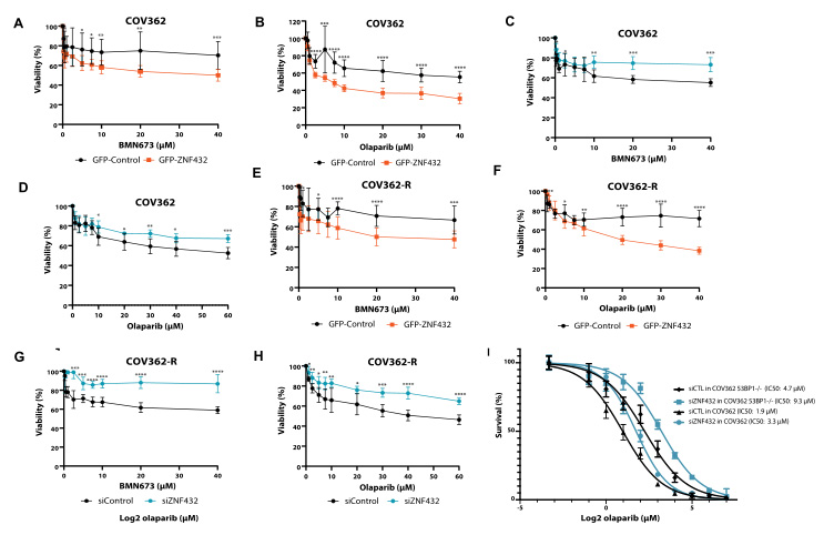
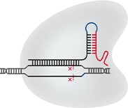
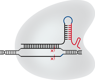
ZNF432 knockout U2OS cell line constructed by Ubigene to carry out a series of experiments, and found that ZNF432 deletion in cancer cells would accelerate DNA repair, lead to the weakening of PARPi effect, and make ovarian cancer cells develop drug resistance, confirming that ZNF432 is a new HR inhibitor, which successfully broadened the new way to study the efficacy of PARPi. View details>>
Regulation of ZNF432 expression affects drug resistance of ovarian cancer cells
SNORD17 Knockout in HepG2 cell line——liver tumors
IF=12.4
Cell death & differentiation
SNORD17 Knockout in HepG2 cell line——liver tumors
IF=12.4 Cell death & differentiation
Abstract:
This study reveals the regulatory role of small nucleolar RNA SNORD17 and p53 pathway in hepatocellular carcinoma, which provides a new potential target for the treatment of hepatocellular carcinoma[5] . The researchers utilized SNORD17 knockout Hep G2 cell line (constructed by Ubigene) as the key cell model. After applied with in vitro and in vivo tests, they found out that in HCC cell lines, the knockout of SNORD17 gene can significantly inhibit cell proliferation, clone formation and G1/S phase transition. View details>>
p53 mediates SNORD17 to affect HCC cell growth
Pik3r1 knockout RAW264.7 cell line - Osteoporosis
IF=11.4
Redox Biology
Pik3r1 knockout RAW264.7 cell line - Osteoporosis
IF=11.4 Redox Biology
Abstract:
This article explores a new mechanism for GSTP1 affecting the osteoclast cell-related bone homeostasis through combining with a large number of in vivo and in vitro experiments, based on the Pik3r1 knockout RAW264.7 cell model constructed by Ubigene. It was the first time to interpret that the cell fate of osteoclasts is determined by S-glutathionylation via a redox-autophagy which is mediated by GSTP1. View details>>
SiNPs induces CASA mechanism
KO cells are often used as key models for studying gene function, disease mechanisms, screening drug targets, etc . Based on the rich gene editing experience, Ubigene has established a stock of over 3500 KO cells, which can meet most scientific research needs. If you would like to get a KO cell line in-stock/customized, please contact us>>>
References:
[1]Zhao C, Ma Y, Zhang M, et al. Polyamine metabolism controls B-to-Z DNA transition to orchestrate DNA sensor cGAS activity[J]. Immunity, 2023, 56(11): 2508-2522. e6.
[2]Wei H, Wang H, Wang G, Qu L, Jiang L, Dai S, Chen X, Zhang Y, Chen Z, Li Y, Guo M, Chen Y. Structures of p53/BCL-2 complex suggest a mechanism for p53 to antagonize BCL-2 activity. Nat Commun. 2023 Jul 18;14(1):4300.
[3]O’Sullivan J, Kothari C, Caron M C, et al. ZNF432 stimulates PARylation and inhibits DNA resection to balance PARPi sensitivity and resistance[J]. Nucleic Acids Research, 2023, 51(20): 11056-11079.
[4]Zhu Y, Zhang Y, Fan Z, et al. Silica Nanoparticles Trigger Chaperone HSPB8‐Assisted Selective Autophagy via TFEB Activation in Hepatocytes[J]. Small, 2023, 19(5): 2204310.
[5]Liang J, Li G, Liao J, et al. Non-coding small nucleolar RNA SNORD17 promotes the progression of hepatocellular carcinoma through a positive feedback loop upon p53 inactivation[J]. Cell Death & Differentiation, 2022, 29(5): 988-1003.

×

Subscribe Us

By subscription, you consent to allow Ubigene Biosciences to store and process the information provided above to deliver the latest news, research spotlight, and promotions. You can unsubscribe from these communications at anytime.
×

Search documentation

Literature:

Name: *

Company: *

Telephone: *

Email:

Notes: After submitting the order, we will contact you as soon as possible.

request now