[Research Highlight] TXNIP positively regulates the autophagy and apoptosis in the rat müller cell of diabetic retinopathy

Diabetic retinopathy (DR) is a microvascular complication of diabetes, threatening the vision of working-age and aged population worldwide, which is preventable [1]. Though various studies have been reported in exploring the mechanism and treatments of DR, there are still many unknowns surrounding the disease which calls for further investigation. DR is a neurovascular disease which damages both the retinal vessels and neural cells [2]. As the primary neuroglia crossing from the outer to the inner layer of neuroretina, müller glia provides nutrition and structural stability, which is vital for the homeostasis of retina [3]. Impairment of müller cell under hyperglycemic condition can certainly induce injury to the neuroretina. Therefore, researches in the relationship between müller glia cell and DR are recommended.
Haocheng Ao from Ophthalmology Center of Sun Yat-sen University published an article "TXNIP regulates the autophagy and apoptosis in the rat muller cell of diabetic retinopathy" in Life Sciences (2020IF:3.647) to investigate the effects of TXNIP on autophagy and apoptosis of Muller cells in diabetic retinopathy rats.
Fig 1
Reverse transcription-quantitative polymerase chain reaction (RT-qPCR) and western blotting were used to measure the expression level of the targets. Clustered regularly interspaced short palindromic repeats/CRISPR-associated 9 (CRISPR/cas9) method was applied for knockout of TXNIP. TdT-mediated dUTP Nick-End Labeling (TUNEL) assay and flow cytometry were utilized to detect the apoptosis. Cell Counting Kit-8 (CCK-8) assay was used to evaluate the cell viability. EdU assay was carried out to measure the cell proliferation ability. Retinal immunohistochemistry, retinal frozen section immunofluorescence as well as the electroretinogram (ERG) recording were implemented to detect the function of the retina.
TXNIP was up-regulated under hyperglycemic condition both in vivo and in vitro. Overexpression of TXNIP activated the autophagy and apoptosis in the rat müller cell. Knockout of TXNIP reduced the autophagy and apoptosis in the rat müller cell under high glucose condition. TXNIP positively regulates autophagy via inhibition of the PI3K/AKT/mTOR signaling pathway. Knockdown of TXNIP improved the visual response to light stimulus of DR.
To confirm the role of TXNIP in rMC-1, researchers designed a lentivirus overexpressing rat gene TXNIP (Fig. 2A, provided by Ubigene). RT-qPCR revealed that TXNIP mRNA level of the overexpression group was significantly higher than that of the control vector group (Fig. 2B). In addition, western blotting showed similar increased expression of TXNIP protein level in overexpression group (Fig. 2C). To evaluate the autophagic flux in rMC-1 under TXNIP overexpressing condition, bafilomycin A1 was applied for blocking autophagic flux. As shown in Fig. 2D, the LC3B-II protein level of TXNIP overexpression group significantly increased with bafilomycin A1, which meant that autophagic flux was enhanced. Besides, the expression of autophagy marker p62 declined while Beclin-1 and Atg12-Atg5 compound increased in the overexpression group. Furthermore, researchers found that the Bcl-2 protein level decreased while Bax and Cleaved Caspase-3 rose in TXNIP overexpression group (Fig. 2E), which suggested that overexpressing TXNIP upregulated apoptosis.
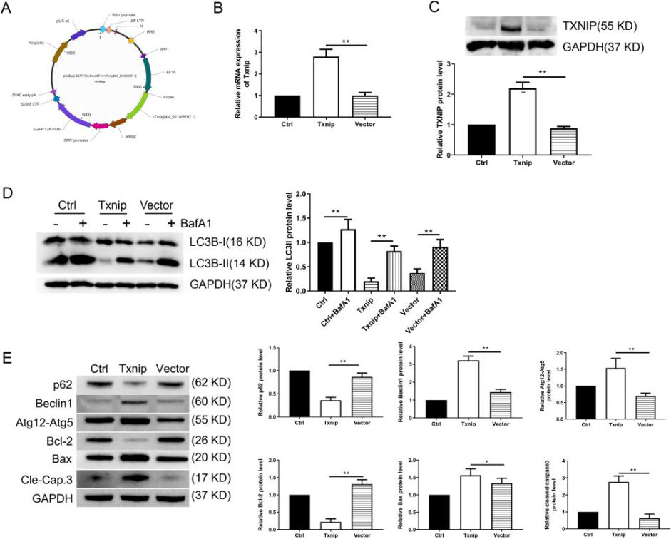
Fig 2
In conclusion, based on the findings of our study, researchers validated the overexpression of TXNIP in vivo and in vitro under hyperglycemic condition, which may serve as a potential biomarker in assessing the severity of diabetic retinopathy. Our study unraveled for the first time that TXNIP positively regulates the autophagy in rat müller cell under high glucose condition by inhibiting the PI3K/AKT/mTOR signaling pathway. Downregulation of TXNIP leads to the reduction of autophagy as well as apoptosis, restoring the cell proliferation and cell viability under high glucose condition, and improves the visual function of diabetic rats. This study sheds light on a potential novel therapeutic approach for DR.
Ubigene provides various types of gene-editing vectors for different species and different virus packaging services, which will easily achieve microbial gene knockout, point mutation and knockin. Vectors and virus offered by Ubigene has been successfully applied in the gene knockout and overexpression on more than 100 cell lines.
Ubigene developed CRISPR-U™ which optimizes eukaryotic cells and animal gene-editing vectors and processes. The efficiency and accuracy are 10x higher than traditional methods. Contact us immediately to know about your research related services!
Reference:
Haocheng Ao, Haichun Li, Xiujuan Zhao, Bingqian Liu, Lin Lu. TXNIP positively regulates the autophagy and apoptosis in the rat müller cell of diabetic retinopathy[J].Life Sciences, 2021: vol 267